Blog
SARS-CoV-2 infection in coronary vessels linked to cardiovascular complications
In a recent study published within the journal Nature Cardiovascular Research, researchers reported that the increased long-term risk of acute cardiovascular complications related to coronavirus disease 2019 (COVID-19) is linked to severe acute respiratory syndrome coronavirus 2 (SARS-CoV-2) infecting coronary vessels and inducing the formation of plaques.
Background
The clinical presentations of COVID-19 have been widely varied, starting from asymptomatic to acute respiratory distress, complications involving multiple organs, and even death.
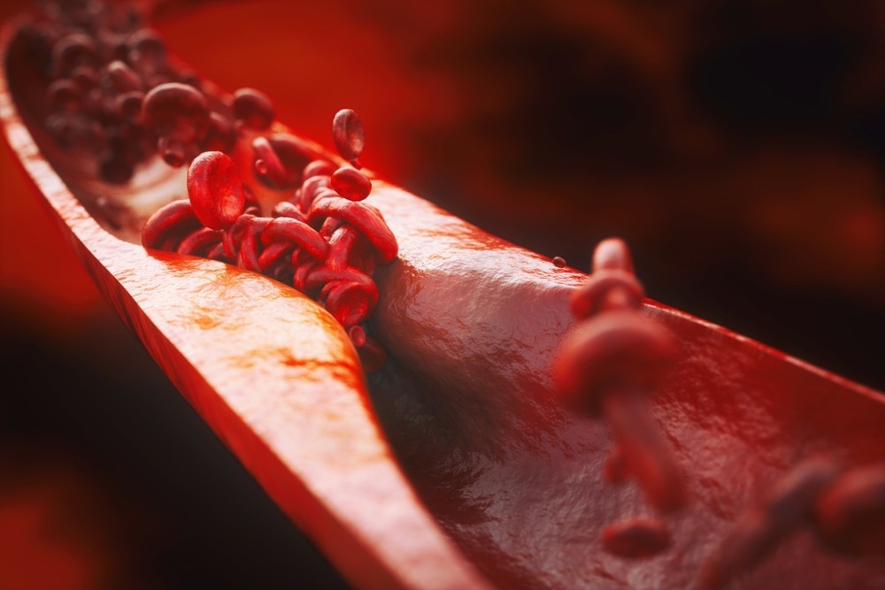
One among the clinical complications of SARS-CoV-2 infections is ischemic cardiovascular events, including stroke and acute myocardial infarctions (AMI), which manifest when atherosclerotic plaques which are chronically inflamed get disrupted.
While strokes and AMIs have been observed within the case of several respiratory infections, reminiscent of those involving influenza virus, the chance of stroke is greater than seven-fold higher for COVID-19 patients.
Moreover, the cytokine storm involving an extreme inflammatory response often related to severe COVID-19 cases is believed to extend the chance of stroke and AMIs. Nevertheless, while the direct infection of lungs and other organs reminiscent of the brain, kidney, adipose tissue, gut, and myocardium by SARS-CoV-2 has been documented, whether the virus directly infects the coronary vasculature and its role in increasing the chance of stroke or AMIs stays poorly understood.
Concerning the study
In the current study, the researchers used coronary autopsy specimens from patients with reverse transcription polymerase chain response (RT-PCR)-confirmed COVID-19 between May 2020 and 2021. The autopsy reports and medical health records provided relevant demographic data and medical history, including the cardiovascular risk aspects and clinical characteristics.
Of the eight patients included within the study, three were diagnosed with acute myocardial ischemia in the course of the hospitalization, one experienced a stroke, and the autopsy results of 4 patients revealed coronary stenosis. Coronary artery sections from autopsy samples were hematoxylin and eosin stained and classified as pathological intimal thickening with macrophage infiltration, fibrocalcific plaque, adaptive intimal thickening, and fibroatheroma by a cardiovascular pathologist. Immunohistochemical staining for CD68+ was also performed.
Ribonucleic acid (RNA) fluorescence in situ hybridization (RNA-FISH) was conducted to detect the viral RNA encoding the SARS-CoV-2 spike protein. Moreover, the antisense strand of the spike protein gene was also probed to substantiate the infection of the coronary vasculature by SARS-CoV-2 because the antisense strand is barely produced when the virus replicates. The cellular localization of the viral RNA was established by identifying the macrophage infiltration of coronary vessels using CD68 probes.
Moreover, a neural network artificial intelligence-based method was used to categorise the perivascular fat and coronary artery wall in each section, given the power SARS-CoV-2 has to contaminate and accumulate viral RNA in adipose tissue.
Nuclear segmentation was also used to quantify the infiltration of the perivascular fat and coronary artery wall with RNAscope probes. Further RNAscope analyses using single-cell RNA sequencing (scRNAseq) datasets of mice and humans were used to detect the potential spread of SARS-CoV-2 to other cells, especially vascular smooth muscle cells.
Foam cells, that are macrophages laden with cholesterol and the buildup of which is indicative of assorted stages of heart problems, were also investigated for SARS-CoV-2 infection, as were macrophages. These cells were also experimentally infected with a modified fluorescent reported virus carrying the isolate from SARS-CoV-2 USA WA1/2020 to know whether the virus could infect plaque macrophages and foam cells.
Results
The findings reported that the viral RNA of SARS-CoV-2 was detected and located to copy within the autopsy samples of coronary vasculature obtained from severe cases of COVID-19. Moreover, the virus showed a stronger tropism for plaque macrophages and arterial lesions than the perivascular fat surrounding the lesions, which also correlated to the degrees of macrophage infiltration.
Moreover, foam cells were more prone to getting infected with SARS-CoV-2 than the opposite macrophage types, and the infection was depending on the neuropilin-1 receptor. The SARS-CoV-2 infection of human vascular carotid explants conducted ex-vivo also showed that the virus stimulated strong inflammatory responses in foam cells and macrophages that were pro-atherogenic. That is believed to exacerbate ischemic cardiovascular complications in severe COVID-19 cases.
Nevertheless, the authors stated that given the small cohort size investigated within the study, which largely consisted of older patients with pre-existing cardiovascular complications, the outcomes can’t be generalized to the younger, healthier populations. Moreover, the findings were based on the strains in circulation within the early phases of the pandemic, and the replication of subsequent SARS-CoV-2 strains within the coronary vasculature can’t be confirmed based on these results.
Conclusions
Overall, the findings suggested that SARS-CoV-2 exhibited a tropism for foam cells and plaque macrophages over the encompassing perivascular fat in coronary vasculature.
Ex-vivo experiments also demonstrated that SARS-CoV-2 infections in macrophages and vascular explants induced strong inflammatory responses and increased cytokine secretion related to cardiac events.
The outcomes indicated that SARS-CoV-2 infections within the coronary vasculature induce plaque formation and increase the chance of cardiovascular complications.
Journal reference:
- Eberhardt, N., Noval, M. G., Kaur, R., Amadori, L., Gildea, M., Sajja, S., Das, D., Cilhoroz, B., Stewart, O. J., Fernandez, D. M., Shamailova, R., Guillen, V., Jangra, S., Schotsaert, M., Newman, J. D., Faries, P., Maldonado, T., Rockman, C., Rapkiewicz, A., & Stapleford, K. A. (2023). SARSCoV2 infection triggers proatherogenic inflammatory responses in human coronary vessels. Nature Cardiovascular Research. doi: https://doi.org/10.1038/s44161023003365 https://www.nature.com/articles/s44161-023-00336-5

