Blog
Examining the cardiac pathology in fatal cases of yellow fever
In a recent study published in eBioMedicine, researchers examined the histopathological features and immunohistochemical and proteomic characteristics of yellow fever (YF)-related myocardial damage.
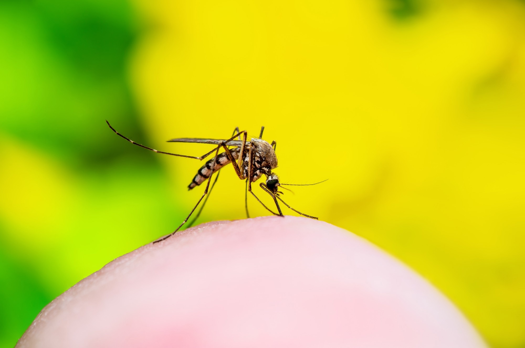
Study: Understanding yellow fever-associated myocardial injury: an autopsy study. Image Credit: nechaevkon/Shutterstock.com
Background
Yellow fever is characterised by a hemorrhagic fever of viral etiology with significant morbidity prevalent in portions of Africa and South America. Heart involvement in yellow fever has been observed in studies, including a high prevalence of bradycardia and electrocardiographic abnormalities, with some data suggesting myocarditis.
Pathology investigations revealed abnormalities similar to hemorrhages, edema, unusual myocarditis, and immunohistochemistry-detectable YF virus (YFV)-antigens within the myocardium. Nevertheless, the pathophysiology of myocardial damage just isn’t clear.
Concerning the study
In the current retrospective autopsy study, researchers described microscopic findings within the cardiac tissues of people decreased on account of yellow fever virus infections.
The study included confirmed yellow fever cases autopsied on the central morgue of São Paulo by the Death Verification Service within the 2017 to 2019 period.
Medical records were reviewed, and the researchers subjected the cardiac tissues to histopathological assessments, immunohistochemistry (IHC) assays, transmission electron microscopy (TEM), and proteomics analyses on endothelial and inflammatory biomarkers.
As well as, quantitative reverse transcription-polymerase chain response (RT-qPCR) was used to quantify yellow fever virus (YFV) ribonucleic acid (RNA).
The team investigated the underlying processes of YF-related cardiac injury and detailed its clinical and pathological characteristics.
Individuals with yellow fever vaccine-related viscerotropic illness (YEL-AVD) who had positive RT-PCR findings for the vaccine virus but negative for the wild-type viral strain were also included. As well as, individuals from the Pathology Department who died on account of cardiovascular illness or sepsis were included as controls.
Patients’ records were looked for clinical and demographic data. Their age, sex (as determined at birth), medical history, cardiovascular and clinical events during hospital admission, interventions, troponin levels, echocardiograms, and electrocardiograms (ECGs) were also obtained.
The researchers performed all autopsies using the Letulle approach. The histology of all samples was assessed by a pathologist specializing in postmortem and infectious diseases pathology and a cardiologist; discrepancies were settled by consensus.
Results
In total, 696 YF cases were documented in São Paulo from 2017 to 2019, and 232 (33%) deaths were reported, amongst whom 73 patients, with a median age of 48 years, were autopsied. Most patients (85%) were men, suffered from hypertension (29%), consumed alcohol (51%) and smoked cigarettes (37%).
The median length of hospital stay was five days and the median interval from symptom onset to death was nine days. All YFV-infected individuals developed shock, required vasopressors, and 4 moreover required inotropes. Supraventricular tachyarrhythmias, the Faget sign, and bradyarrhythmias were reported in 21%, 11%, and 6.8% of people.
All individuals died from refractory-type shock with hemorrhagic and septic components brought on by coagulopathy, severe renal damage, hepatic failure, and hepatic encephalopathy.
On account of YFV infections, the researchers discovered a major incidence of hemorrhages, interstitial edema, endothelial abnormalities, myocardial fibrosis, mononuclear myocarditis, and hypertrophied and necrosed cardiomyocytes.
Fibrosis and enlarged cardiomyocytes were observed within the myocardial tissues of 68 individuals (93%), endothelial changes in 67 individuals (92%), fiber necrosis amongst 50 individuals (69%), myocarditis amongst nine (12%), and secondary myocardial inflammation amongst five individuals (7.0%). Myocarditis was present in 4 of 5 individuals affected by 17DD vaccine-related viscerotropic illness.
Endothelial fibrinoid necrosis, hemorrhages, and edema were observed within the cardiac conduction system. YFV components present in the myocardial tissues include virus-like particles (via electron microscopy in endothelial cell cytoplasm), YFV-RNA via RT-qPCR, and YFV antigens via immunohistochemistry (in inflammatory and endothelial cell cytoplasm), in a single, 66, and 24 cases, respectively.
IHC examination revealed CD68-expressing inflammatory cells within the interstitium and yellow fever virus antigens within the inflammatory and endothelial cells. In 96% of cardiac specimens, YFV-RNA was found to be positive.
Individuals with YFV infections showed increased levels of assorted endothelial and inflammatory biomarkers in comparison with controls, in addition to elevated interferon-gamma (IFN-γ)-induced protein 10 (IP-10) amongst severely infected individuals in comparison with sepsis and controls within the proteomic evaluation.
Implications
Overall, the study findings showed that myocardial damage is a typical complication of severe YF, and various clinical characteristics and multiple processes characterize it.
This injury may end in direct yellow fever virus-mediated injury, local and systemic inflammation, secondary fungal or bacterial sepsis, cardiomyopathy, and endothelial damage.
The study findings indicated that physicians should perform complete cardiovascular examinations on patients with YF, allowing for early therapeutic and supportive actions.
The findings may potentially aid in developing novel biomarkers for YF myocardial damage. Yellow fever will be prevented with a really effective vaccination (17DD), but its stockpile and treatment arsenal are limited.
Furthermore, the 17DD vaccine’s viral strain can result in end-organ dysfunctions, including liver, brain, and heart failure. To help in YF prevention, globally coordinated efforts are required, and pathology investigations are critical to understanding the damage processes in diverse organs.

