Blog
A review of the neurological imaging techniques in dementia diagnosis
In a recent review published by Molecular Psychiatry, researchers reviewed existing data on the contribution of neuroimaging modalities similar to magnetic resonance imaging (MRI) and positron emission tomography imaging (PET) in dementia diagnosis.
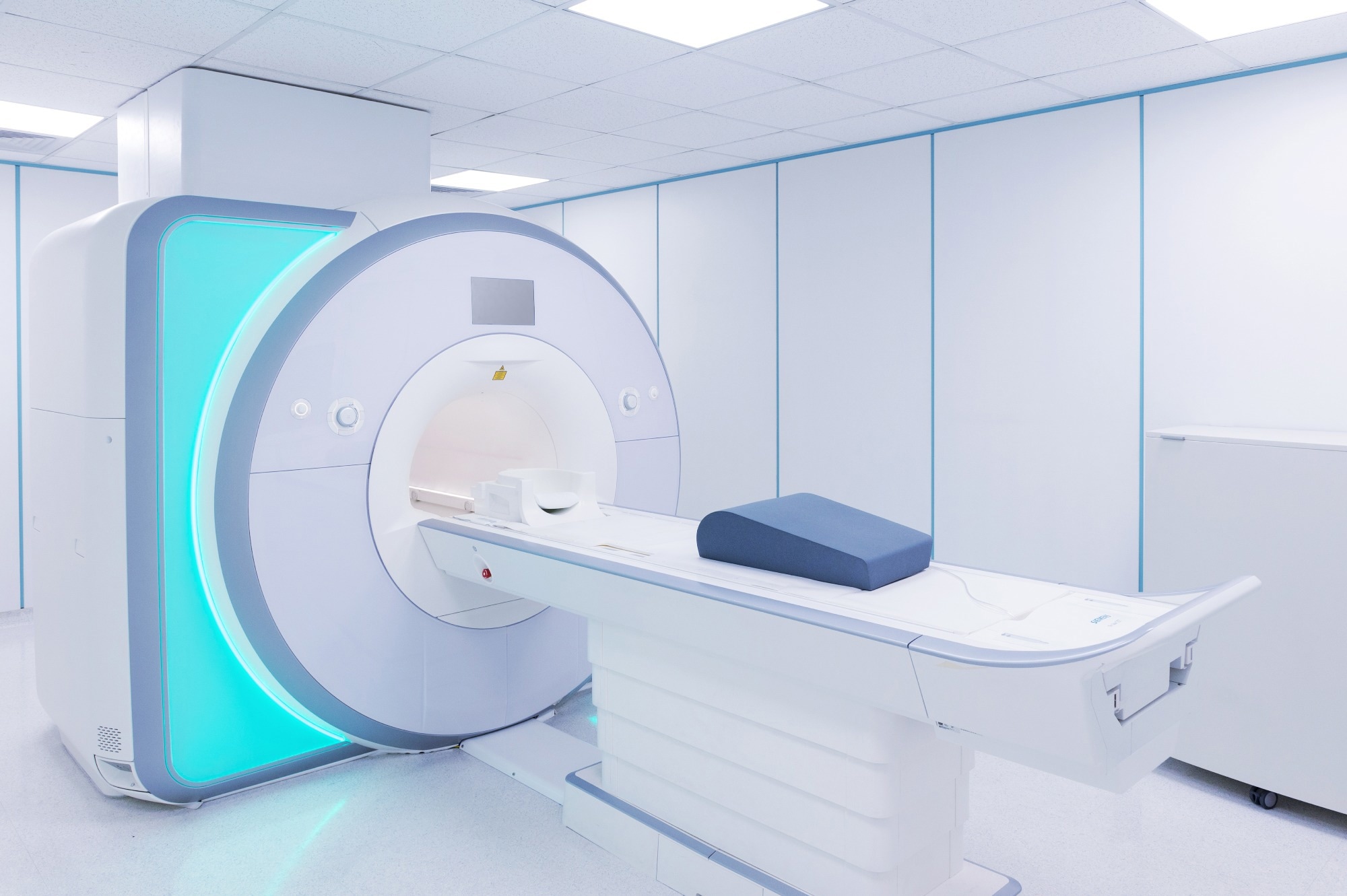
Study: Using neuroimaging techniques within the early and differential diagnosis of dementia. Image Credit: KaliAntye/Shutterstock.com
Background
Dementia has resulted in disability, cognitive impairment, and mortality worldwide. It necessitates early detection and precise treatment for prevalent kinds similar to Vascular dementia, Alzheimer’s disease (AD), Frontotemporal dementia (FTD), and Lewy Body Dementia (LBD).
Molecular and structural imaging assists in understanding the etiology of neurodegenerative-type dementias, allowing for higher clinical care and therapies.
Concerning the review
In the current review, researchers presented the role of neuroimaging in dementia diagnosis.
Magnetic resonance imaging to evaluate neurodegeneration
Structural magnetic resonance imaging is a crucial diagnostic technique for dementia based on atrophy, white matter abnormalities, and cerebrovascular illness.
It might aid within the identification of disorders similar to space-occupying lesions, stroke, and normal-pressure hydrocephalus, and the differentiation of dementia from other illnesses. In Alzheimer’s disease, temporal lobe atrophy is widespread, with localized alterations within the hippocampus.
Volumetric MRI measures pathological alterations with techniques similar to voxel-based morphometry and cortical thickness evaluation.
Volumetric MRI alterations, then again, are insensitive in early prodromal dementia phases and customarily correspond with established neurodegenerative processes. Serial MRI has been utilized to boost dementia differential diagnosis and is regularly employed as a secondary end result measure in treatment studies.
Diffusion tensor imaging (DTI) is an MRI method that evaluates the direction and integrity of white matter pathways within the brain by evaluating aspects related to water molecule diffusion. DTI data can also be employed in analytical approaches like tractography. Recent MRI investigations have revealed physiologically plausible variations between dementia subtypes and predicted the transition from MCI to dementia.
Blood flow could also be measured using injectable contrast agents or arterial spin labelling (ASL) in functional MRI, and variations in blood flow correlate with FDG-PET results.
Magnetic resonance spectroscopy, magnetoencephalography, and neurite orientation dispersion and density imaging (NODDI) are examples of MR-based advances in neuroimaging.
F-FLUORODEOXYGLUCOSE (FDG) PET imaging in identifying neurodegenerative diseases
FDG-PET, which measures the local brain metabolic rate of glucose consumption, is often used to diagnose Alzheimer’s disease and other forms of dementia. It has a high specificity in identifying roughly half of the individuals with behavioral variant FTD (bvFTD) who’re undetectable by MRI, allowing the exclusion of psychiatric and other neurodegenerative illnesses.
Hypometabolism on FDG-PET could be found a minimum of 10 years before symptoms start in hereditary sorts of FTD, and the severity of symptoms corresponds with more broad regions of hypometabolism on FDG-PET. Nevertheless, FDG-PET needs to be utilized together with a history and other available examinations.
Neuroimaging is a well-established neurodegenerative marker, including brain dopamine transporter imaging, cardiac sympathetic nerve imaging (MIBG), and single-photon emission computed tomography (SPECT).
Amyloid PET is a big imaging technique within the initial and particular diagnosis of Alzheimer’s disease, notably within the diagnosis of young-onset Alzheimer’s disease, and it distinguishes it from other dementias.
Nevertheless, it has just a few limitations, including the proven fact that it shouldn’t be correlated with symptomatic onset or disease severity, that it cannot predict dementia onset, and that more research is required for older individuals as a result of the prevalence of amyloid pathology amongst a big fraction of the older and cognitively unimpaired population.
Tau protein PET imaging, which uses ligand molecules that bind with neurofibrillary tangles, has been authorized by the Food and Drug Administration (FDA) for in vivo tau evaluation in individuals with Alzheimer’s disease.
It could not, nevertheless, be appropriate for non-AD tauopathies. Aside from amyloid and tau protein, other essential PET ligands include 11C-UCBJ, 18F-SynVesT-1, and 11C -PK11195.
Conclusions
Overall, the review findings emphasized the importance of neuroimaging in dementia diagnosis, with structural MRI being regularly employed in routine clinical practices. Nevertheless, its sensitivity for early diagnosis and differential identification of dementia subgroups is sort of poor.
FDG-PET imaging has high specificity and sensitivity for FTD and AD, but PET with tau and amyloid protein ligands can enhance the differentiation of Alzheimer’s disease-related and non- Alzheimer’s disease dementias, including identification at prodromal phases.
Dopaminergic-type imaging might help in diagnosing LBD, although there are currently no validated tracers for TAR DNA-binding protein 43 (TDP-43) or alpha-synuclein imaging.
Experts agree on three approaches for clinical dementia diagnosis: Amyloid protein PET or cerebrospinal fluid (CSF) examination within the case of suspected AD; FDG-PET if the primary workup reveals non- Alzheimer’s disease dementia; and [(123)I]N-omega-fluoropropyl-2beta-carbomethoxy-3beta-{4-iodophenyl}nortropane ([(123)I]FP-CIT) SPECT imaging or MIBG in cases of cognitive deficits related to movement-related conditions.
Neuroimaging of the brain is increasingly being utilized to stratify clinical trial participants and assess treatment response in studies of disease-modifying pharmaceutical agents.
The amyloid/tau/neurodegeneration (ATN) framework for Alzheimer’s disease diagnosis, which specifies the existence of Alzheimer’s disease pathology within the preclinical in addition to prodromal phases, is critical to applying disease-modifying drugs (DMDs) in clinical care.

